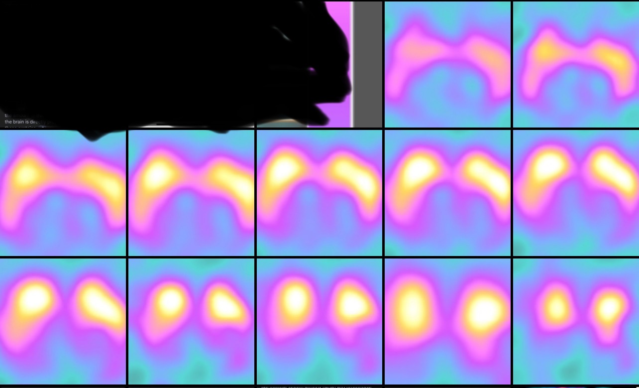
Datscan pictures below are normal supposedly I know I ain’t a neurologist but to untrained eye these definitely don’t look aysemtrical possibly do in shape but certainly not activity.
I was diagnosed clinical probable Parkinson’s by 3 dif neurologists including MDS. Got shown nurse started meds ect and given newly diagnosed pack (meds were working now weaned off due to being told my datscan results are robust and has came back normal for a late 30 yr old man so meds must have been a placebo effect)
Anyone able to compare if they pref in late 30s
So what I know of a datscan (happy to be corrected)
In a DaTscan, the radioactive tracer, loflupane (123l), outlines the dopamine transporters (DATs) in the brain, while the “inside” refers to the activity or uptake of the tracer within those transporters. Areas showing tracer uptake should display SYMMETRICAL ACTIVITY,
the tracer should be similar on
both sides of the brain.
research also suggests that starting at approximately 30% but upto if age 49/50 which still classed as young onset you can lose all way upto 80% of dopamine-producing neurons by the time motor symptoms become noticeable but it does only take 30% loss to notice motor problems.
So basically A Datscan image showing reduced activity in the putamen on one side compared to the other, while the putamen is still visible, suggests a potential dopamine deficiency, which can be a sign of Parkinson’s disease or other movement disorders. This asymmetry in dopamine transporter activity is a key feature that DaTSCAN can highlight, aiding in diagnosis. Especially if the less active side is in line with the clinical examination ( in my case my left side effected so right side of scan/left side on pictures)